Anatomy Of Chest And Heart / The Lungs Position Structure Teachmeanatomy : Heart, organ that serves as a pump to circulate the blood.. The loose fitting superficial part of this sac is the fibrous pericardium. It is located in the middle cavity of the chest, between the lungs. Normal thoracic ct (lungs, pleura, mediastinum and heart). The right atrium and left atrium receive blood returning from the systemic and pulmonary circuits. Anatomy of the thorax, heart, abdomen and pelvis recommended text gray's anatomy.
Compression of the heart and great vessels may cause murmurs. Yen ho, phd frcpath fesc fhea royal brompton hospital. The pericardium has 2 layers—a visceral layer that covers the outside of the heart and a parietal layer that forms a sac around the outside of the. Normal thoracic ct (lungs, pleura, mediastinum and heart). The conducting system of the heart.
Anatomy Of The Heart And Lungs Diagnosis 101 from diagnosis101.welchallyn.com Current imaging techniques can show in exquisite detail the heart in its anatomical position inside the living patient's chest and. Radiological anatomy of the lungs, mediastinal lymph nodes, trachea, bronchi, pleural cavity, heart and pulmonary vessels. Therefore, the funnel chest is also called 'cobbler chest'. It is located in the middle cavity of the chest, between the lungs. It consist of four chambers, four valves, arteries (named as coronary arteries), and the conduction system. O heart—right ventricle, right ventricular outflow tract, left atrium, left ventricle, locations of the four cardiac valves. This is a thin protective coating that surrounds the other parts. If we want to understand how the heart performs its vital role, we will first have to look at its structure, i.e., cardiac anatomy.
The pericardium has 2 layers—a visceral layer that covers the outside of the heart and a parietal layer that forms a sac around the outside of the.
Anatomy of the chest wall. ■ describe the anatomical relationships of various organs in the chest. The loose fitting superficial part of this sac is the fibrous pericardium. When a patient flexes the neck forward, the prominent process is usually that of the 7th cervical. Learn about and chest heart anatomy with free interactive flashcards. Normal anatomy of the thorax on labeled chest ct: The heart is a muscular organ in most animals, which pumps blood through the blood vessels of the circulatory system. Located between the lungs in the middle of the chest, the heart pumps blood through the network of arteries and veins known as the cardiovascular system. Learn more about the heart in this article. Normal thoracic ct (lungs, pleura, mediastinum and heart). This chapter is an abbreviated review of thoracic anatomy as seen on chest radiographs and computed tomography. Traditionally, the heart is described as having left heart and right heart chambers. This image shows the four chambers of the heart and the direction that blood flows through the heart.
Heart functionally can be separated in left and right side. The heart and circulatory system make up your cardiovascular system. Learn about the organ's amazing power and the functions of its many parts. Yen ho, phd frcpath fesc fhea royal brompton hospital. Compression of the heart and great vessels may cause murmurs.
12 General Anatomy Of The Heart Radiology Key from i1.wp.com Therefore, the funnel chest is also called 'cobbler chest'. Yen ho, phd frcpath fesc fhea royal brompton hospital. When a patient flexes the neck forward, the prominent process is usually that of the 7th cervical. Heart anatomy focuses on the structure and function of the heart. Your heart does a lot of work to keep the body going. The heart is located in the center of the chest with its apex toward the left. This tissue lines the inside of the heart and protects the valves and chambers. ■ describe the anatomical relationships of various organs in the chest.
■ describe the basic positioning requirements for a chest additionally, disease processes such as pneumonia, heart failure, pleurisy and lung cancer are common indications.
■ identify the basic anatomy seen on a chest radiograph. Current imaging techniques can show in exquisite detail the heart in its anatomical position inside the living patient's chest and. The loose fitting superficial part of this sac is the fibrous pericardium. Heart is a muscular organ sited in the mediastinum. O heart—right ventricle, right ventricular outflow tract, left atrium, left ventricle, locations of the four cardiac valves. Learn about and chest heart anatomy with free interactive flashcards. The heart is located in the center of the chest with its apex toward the left. A good radiologist knows the anatomy, so don't skip this chapter! Learn more about the heart in this article. Your heart is located between your lungs in the middle of your chest, behind and slightly to the left of your breastbone. Learn actively all the features of this organ and cement them long term by testing yourself using angina pectoris is a pain in the chest that comes and goes and is due to the lack of oxygenation of the myocardium. Stable angina is the most common. It is located in the middle cavity of the chest, between the lungs.
Heart functionally can be separated in left and right side. The heart is a muscular organ in most animals, which pumps blood through the blood vessels of the circulatory system. Learn more about the heart in this article. If we want to understand how the heart performs its vital role, we will first have to look at its structure, i.e., cardiac anatomy. Narrowed coronary arteries cause predictable chest pain or discomfort with exertion.
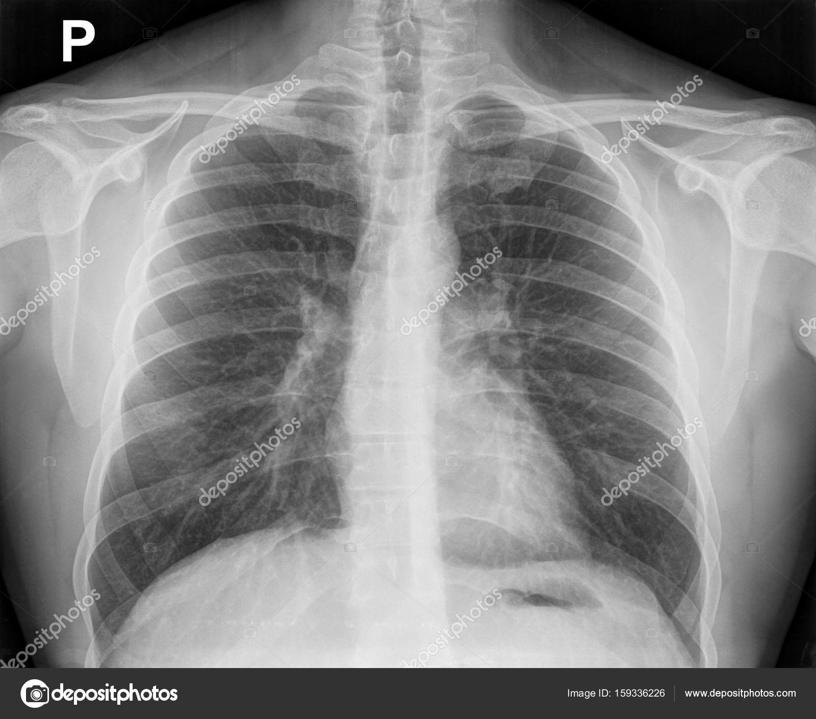
X Ray Image Of The Chest Showing The Internal Anatomy Of The Rib Stock Photo Image By C Gubernat 159336226 from st3.depositphotos.com When a patient flexes the neck forward, the prominent process is usually that of the 7th cervical. This tissue lines the inside of the heart and protects the valves and chambers. Therefore, the funnel chest is also called 'cobbler chest'. Yen ho, phd frcpath fesc fhea royal brompton hospital. Traditionally, the heart is described as having left heart and right heart chambers. This is a thin protective coating that surrounds the other parts. Normal thoracic ct (lungs, pleura, mediastinum and heart). The heart is a muscular organ that pumps blood throughout the body.
Heart is a muscular organ sited in the mediastinum.
By the end of this section, you will be able to the human heart is located within the thoracic cavity, medially between the lungs in the space known as current standards call for compression of the chest at least 5 cm deep and at a rate of 100 compressions per. Radiological anatomy of the lungs, mediastinal lymph nodes, trachea, bronchi, pleural cavity, heart and pulmonary vessels. Related online courses on physioplus. Your heart does a lot of work to keep the body going. The heart is a muscular organ in most animals, which pumps blood through the blood vessels of the circulatory system. Yen ho, phd frcpath fesc fhea royal brompton hospital. Heart dissection gcse a level biology neet practical skills. Narrowed coronary arteries cause predictable chest pain or discomfort with exertion. Our picks for anatomy of the heart and blood vessels. Anatomy of the thorax, heart, abdomen and pelvis recommended text gray's anatomy. The right atrium and left atrium receive blood returning from the systemic and pulmonary circuits. This amazing muscle produces electrical impulses that cause the heart to contract, pumping blood throughout the body. Stable angina is the most common.
Your heart is in the center of your chest, near your lungs anatomy of chest. Heart is a muscular organ sited in the mediastinum.


0 Komentar